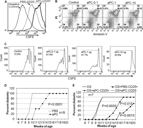
FIGURE 5.
aPC-stimulated Tregs inhibit T cells proliferation and prevent adoptive transferred diabetes in NOD.SCID mice. A, CD4+CD25− T cells (1 × 10⁶ cells/ml) from spleens of NOD mice were labeled with carboxyfluorescein succinimidyl ester and incubated for 72 h in anti-CD3 and anti-CD28 antibody-coated plates either alone (negative control) or with CD4+CD25+ T cells (1:1) from aPC- or PBS-treated mice. Cell proliferation was analyzed by flow cytometry. B, survival/apoptosis of Tregs from mouse spleens in response to aPC (0, 0.1, 1, and 10 μg/ml) for 24 h, detected by annexin V with propidium iodide (PI) staining using flow cytometry. C, proliferation of CD4+CD25− cells (4 × 10⁶ cells/ml) from diabetic NOD mice in response to aPC (0. 0.1, 1, and 10 μg/ml) for 72 h. Cells were stimulated with anti-CD3 and anti-CD28 antibodies, and proliferation was detected by flow cytometry. Data were analyzed with FlowJo software, and representative histogram plots are from triplicate wells of two independent experiments. D, diabetes incidence in NOD.SCID mice after adoptive transfer of spleen cells (1 × 10⁷ cells/mouse) from 26-week-old PBS- or aPC-treated NOD mice. Data are expressed as mean ± S.E. (n = 9). E, diabetes incidence in NOD.SCID mice after adoptive co-transfer of total spleen cells from recently diabetic mice (DS) with splenic CD4+CD25+ or CD4+CD25− cells from 10-week aPC-treated NOD mice or CD4+CD25+ from control mice. DS cells were injected i.v. alone (1 × 10⁷) or mixed (5:1) with CD4+CD25+ cells or CD4+CD25− cells (DS + aPC-CD25−). Data are expressed as mean ± S.E. (n = 7).

