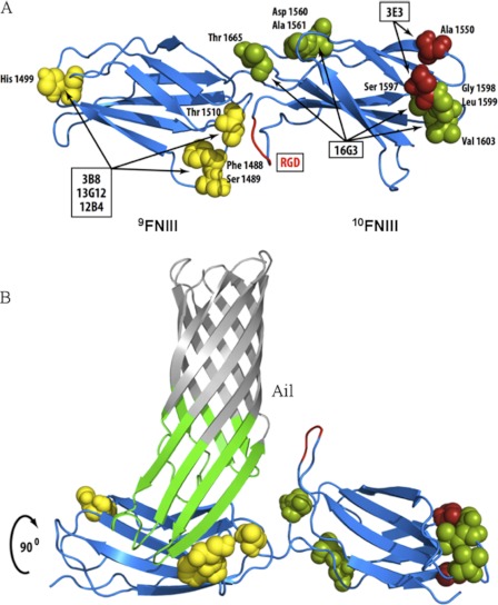
FIGURE 3.
Defining binding sites of mAbs that map to 9–10FNIII. A, based on unique residues in Fn from five different species and the reactivity of each mAb, potential interaction residues in 9–10FNIII are proposed (see also supplemental Fig. S5). B, using OmpX of E. coli as an Ail homolog (62), a model of Ail binding to 9FNIII in a region overlapping the synergy region (51) but exposing the RGD domain is proposed. Repeats 7–10FNIII were structurally determined by Leahy et al. (46).

