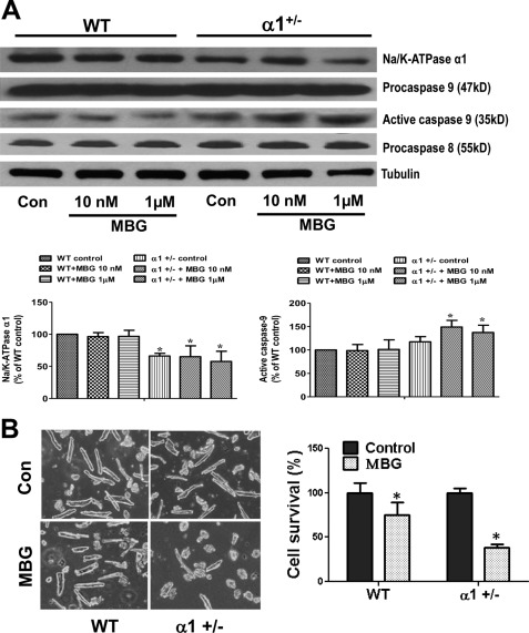
FIGURE 2.
Na/K-ATPase reduction potentiates MBG-induced activation of caspase 9 and cardiac cell death. A, cardiac myocytes were isolated from both WT and α1+/− mice and were treated with different concentration of MBG for 24 h. The cell lysates were collected in RIPA buffer to probe for Na/K-ATPase α1, caspase 9, and caspase 8 using Western blot. Tubulin was used for loading control (Con). The 35-kDa band of caspase 9 indicates the activation of procaspase 9 (a 47-kDa protein). B, isolated adult cardiac myocytes were treated with 1 μm MBG for 24 h. Live cells (rod-shaped) and dead cells (round-shaped) were counted under a microscope (20×) for each group. The left panel shows the representative images of adult myocytes of control and MBG-treated cells. The right panel is the quantification data from three different preparations. *, p < 0.05, control versus MBG treatment.

