FIGURE 6.
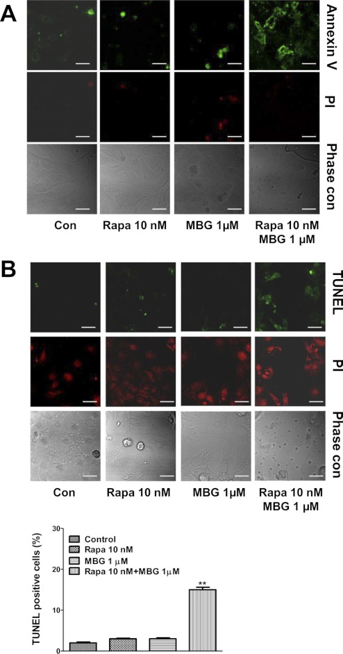
MBG induces cardiac myocyte apoptosis in the presence of rapamycin. Rat neonatal cardiac myocytes were serum-starved for 48 h and then treated with MBG 1 μmol/liter alone or in combination with 10 nmol/liter rapamycin (Rapa) for 72 h. Cell apoptosis was assayed by annexin V staining and TUNEL assay. A, a representative image of cell apoptosis measured by annexin V staining. The green fluorescence of annexin V staining on cell membrane indicates early stage apoptotic cells. The red fluorescence of PI indicates necrosis or later stages of apoptosis. B, TUNEL assay. Apoptotic cells are indicated by green fluorescence-stained nuclei. PI was used for counterstaining. Quantification data were determined by the percentage of TUNEL-positive nuclei. **, p < 0.01 versus control. The scale bar represents 20 μm. Con, control.

