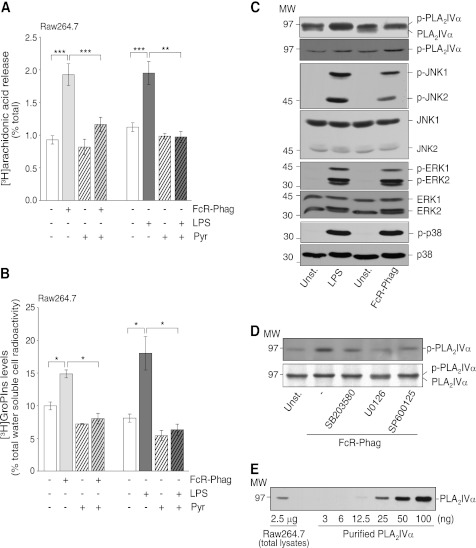
FIGURE 1.
FcR-mediated phagocytosis and LPS activate phosphorylation of PLA2IVα, MAPKs, and stress kinases in Raw264.7 cells. Cells without (−) or with (+) a 15-min preincubation with 0.5 μm pyrrophenone (Pyr) were stimulated by FcR-mediated phagocytosis (FcR-Phag) or by 20 μg/ml LPS from E. coli for a further 30 and 45 min, respectively, at 37 °C (see “Experimental Procedures”). A, [3H]arachidonic acid release (percentage of total [3H]arachidonic acid cell content). Data are means ± S.E. of 10 independent experiments for phagocytosis, each performed in triplicate, and of four independent experiments for LPS stimulation, each performed in duplicate. ***, p < 0.001; **, p < 0.02 (Student's t test). B, intracellular [3H]GroPIns production (percentage of total [3H]inositol-labeled water-soluble metabolites). Data are means ± S.E. of at least five independent experiments, each performed in duplicate. *, p < 0.05 (Student's t test). C, representative Western blotting showing PLA2IVα phosphorylation by gel shift or by phosphorylated PLA2IVα, and phosphorylation (p-) and total levels of JNK, ERK1/2, and p38 in unstimulated cells (Unst.), after 30 min of FcR-mediated phagocytosis or of 20 μg/ml LPS treatments (see “Experimental Procedures”). D, representative Western blotting showing phosphorylated PLA2IVα in unstimulated cells (Unst.) or after 30 min of FcR-mediated phagocytosis in the absence (−) or presence of 15-min preincubation with 10 μm of the indicated kinase inhibitors. E, representative Western blotting of 2.5 μg total Raw264.7 cell lysate and increasing amounts of purified PLA2IVα protein (from 3 to 100 ng, as indicated). MW, molecular weight marker (kDa).

