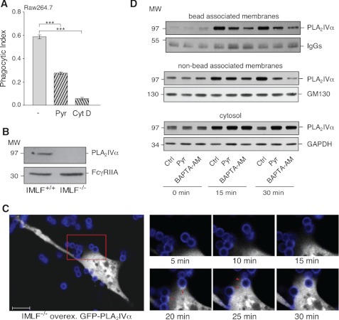
FIGURE 2.
PLA2IVα is a component of the phagocytic machinery formed consequent to FcR activation. Raw264.7 cells were stimulated by FcR-mediated phagocytosis of IgG-opsonized latex beads for 30 min at 37 °C, and particle uptake/binding was quantified. A, quantification of phagocytosed beads by fluorescence microscopy. Phagocytosis was performed in the absence (−) and presence of a 15-min pretreatment with 0.5 μm pyrrophenone (Pyr) or 10 μm cytochalasin D (Cyt D). Data are expressed as phagocytic index and represent means ± S.E. of eight independent experiments. ***, p < 0.001 (Student's t test). B, representative Western blotting showing expression of endogenous PLA2IVα (top) and overexpressed FcγR (bottom) in IMLF+/+ and IMLF−/− cells. C, time-lapse microscopy images showing PLA2IVα localization during FcR-mediated phagocytosis. IMLF−/− cells were grown in live cell imaging dishes and transfected with FcγR and GFP-PLA2IVα and then used in the phagocytosis assay in a thermostated chamber under the microscope. FcγR-mediated phagocytosis was followed under fluorescence microscopy. The large panel (left) shows a single IMLF−/− cell transfected with GFP-PLA2IVα (white) in the presence of Alexa546-labeled IgG-opsonized beads (blue). Scale bar, 10 μm. The smaller panels (right) are higher magnifications of the red square outlined in the larger panel, as frames were collected at the times indicated. The arrowheads (20–25 min) indicate GFP-PLA2IVα localization at the phagocytic cup. D, localization of endogenous PLA2IVα during FcR-mediated phagocytosis in Raw264.7 cells. Phagocytosis was performed in the absence (Ctrl) and presence of a 15-min pretreatment with 0.5 μm pyrrophenone (Pyr) or a 5-min pretreatment with 10 μm BAPTA-AM (incubated in the last minutes of opsonized bead binding). At the indicated times (0, 15, and 30 min), phagocytosis was terminated, and the nascent phagosomes (bead-associated membranes), total cell membranes (nonbead-associated membranes), and cytosolic fractions (cytosol) were recovered and subjected to Western blotting (see “Experimental Procedures”). Data are representative of three independent experiments.

