FIGURE 3.
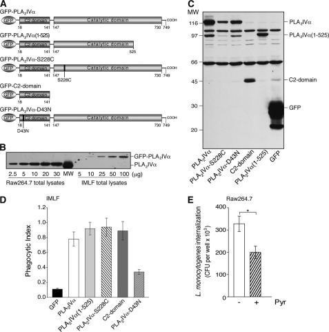
PLA2IVα enzymatic activity is not necessary for productive FcR-mediated phagocytosis. A, schematic representation of GFP-PLA2IVα and PLA2IVα deletion and point mutants. GFP-PLA2IVα is characterized by an N-terminal C2 domain and a C-terminal catalytic domain. The GFP-PLA2IVα-C2-domain, the deleted mutant GFP-PLA2IVα(1–525), and the point mutant GFP-PLA2IVα-S228C have no catalytic activity, but they translocate to membranes under agonist stimulation (18, 36, 55). The point mutant GFP-PLA2IVα-D43N cannot translocate to membranes under cell stimulation (12). B, representative Western blotting of 2.5–30 and 5–100 μg total lysates from Raw264.7 and IMLF−/− cells overexpressing GFP-PLA2IVα, respectively. IMLF−/− transfection efficiency was 30%, as verified by FACS analysis, independent of transfection construct, and the estimated total protein was ∼520 and ∼350 pg/cell for Raw264.7 and IMLF−/− cells, respectively. The PLA2IVα protein content was comparable between 3 μg of Raw264.7 cell lysate and 100 μg of IMLF−/− cell lysate. Thus, assuming uniform overexpression of GFP-PLA2IVα and taking into account the efficiency of transfection, the protein levels reached in PLA2IVα-transfected IMLF−/− cells was one-tenth that of the endogenous level in Raw264.7 cells. C, representative Western blotting of transfection levels of GFP and GFP-PLA2IVα (wild-type and mutants, as indicated) in IMLF−/− cells, followed using an anti-GFP antibody (see “Experimental Procedures”). Data are representative of at least four independent experiments. D, quantification by morphological analysis of particle uptake (phagocytic index) in IMLF−/− cells transfected with FcγR ± GFP or with different GFP-PLA2 constructs (as indicated). Data represent means ± S.E. of at least four independent quantifications. E, internalization of L. monocytogenes in Raw264.7 cells. Cells were infected with bacteria (multiplicity of infection of 10) for 1 h at 37 °C, and internalization was quantified using the gentamicin protection assay (see “Experimental Procedures”) in the absence (−) and presence (+) of 0.5 μm pyrrophenone (30-min pretreatment), and expressed as colony forming units per well. In control proliferation assays, pyrrophenone did not affect L. monocytogenes growth (up to 10 h of pyrrophenone treatment). Data are means ± S.D. of nine independent experiments, each performed in triplicate. *, p < 0.05 (Student's t test).

