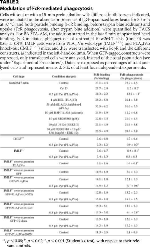
TABLE 2.
Modulation of FcR-mediated phagocytosis
Cells without or with a 15-min preincubation with different inhibitors, as indicated, were incubated in the absence or presence of IgG-opsonized latex beads for 30 min at 37 °C, and both particle binding (FcR binding, before trypan blue addition) and uptake (FcR phagocytosis, after trypan blue addition) were quantified by FACS analysis. For BAPTA-AM, the addition started in the last 5 min of opsonized bead binding. FcR-mediated phagocytosis of untreated Raw264.7 cells (time 0) was 0.65 ± 0.4%. IMLF cells were from PLA2IVα wild-type (IMLF+/+) and PLA2IVα knock-out (IMLF−/−) mice, and they were transfected with FcγR and the different constructs, as indicated in the left-hand column. When GFP-tagged constructs were expressed, only transfected cells were analyzed, instead of the total population (see under “Experimental Procedures”). Data are expressed as percentages of total analyzed cells and represent means ± S.E. of at least four independent experiments.
a, p < 0.05; b, p < 0.02; c, p < 0.001 (Student's t-test), with respect to their relevant controls.

