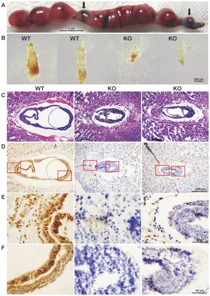
Figure 2. Morphology and histology of Cul4b null embryos.
(A) Uterus excised from pregnant female at 12.5 dpc. Arrows indicate absorbed embryos. (B) Photomicrographs of E7.5 embryos dissected from surrounding deciduas tissue of the same uterus. The two embryos on the left appear normal in size and morphology and the two on the right were much smaller and were partially deteriorated. The bar represents 200 µm. (C) H&E staining of paraffin sections of wild-type and Cul4b null embryos at 7.5 dpc. Littermate embryos at 7.5 dpc were paraffin embedded and cross sectioned together with their surrounding deciduas. The genotype of each embryo was determined by immunohistochemistry using an anti-Cul4b antibody, as shown below. (D) Immunohistochemistry of paraffin sections of wild-type and Cul4b null embryos at 7.5 dpc with an anti-Cul4b antibody. (E–F) Photomicrographs with higher magnification of the stained section shown in (D).

