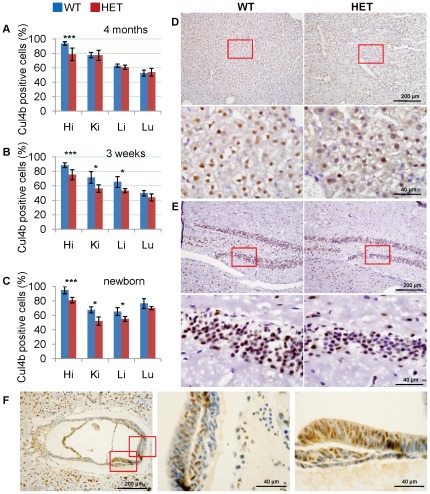
Figure 6. Characterization of X chromosome inactivation by Cul4b expression in heterozygous mice.
(A–C) Percentages of cells positive for Cul4b of Cul4b heterozygous mice and littermate wild-type female controls at 4 months (A), 3 weeks (B) and newborn (C). More than 2,000 cells of each tissue were scored. Hi, hippocampus; Ki, kidney; Li, liver; Lu, lung. Data were presented as mean±SD. *: p<0.05; **: p<0.01; ***: p<0.001. (D–E) Representative images of liver (D) and hippocampus (E) at 3 weeks stained with an antibody against Cul4b. Sections were counterstained with haematoxylin. Lower panels are the higher magnification of the upper panels. (F) Immunohistochemistry of paraffin sections of Cul4b heterozygous embryos at 7.5 dpc with an anti-Cul4b antibody. Embryos at 7.5 dpc were paraffin embedded and cross sectioned together with their surrounding deciduas. Middle and right panels are the higher magnification of the left panel.

