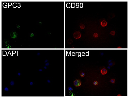
Figure 7. Double immunofluorescence staining of CD90 and GPC3 in sorted PLC CD90+GPC3+ cells.
The sorted cells were stained with fluorescein-conjugated anti-CD90 and anti-GPC3 antibodies. Nuclei were counterstained by DAPI. The merge image showed the expression of CD90 and GPC3 in both cytoplasm and cell membrane.

