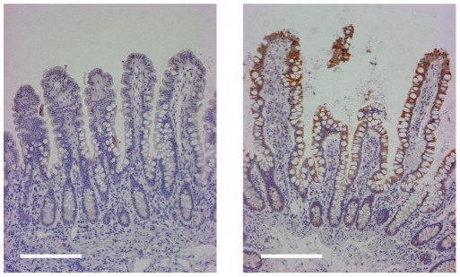
Figure 4. Immunohistochemical localization of FOLH1 in disease unaffected ileal mucosa from the proximal margin of resected ileum from an ileal CD subject (left panel) and a control non-IBD subject.
The more prominent FOLH1 staining in the ileal CD sample is localized to the villous epithelium. Magnification is 100×. Bar is 200 µm.

