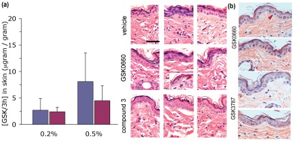
Figure 4. Absence of inflammatory changes induced by PPAR β/δ antagonists in skin after topical application.
(a) C57Bl/6j wild type mice were treated with ointments containing GSK0660 or compound 3 h applied twice daily to shaved dorsal skin for one week. Mice were sacrificed 1 h after the last ointment application and skin tissue processed for H&E based histology and mass spectrometry, as described in Methods. Data shown represent average ± s.d. of n = 3 mice per data point (left) treated with GSK (blue columns) or compound 3 h (red). Representative histology sections of all treated mice are shown on right. The inset in the middle panel shows a section of GSK0660-treated epidermis showing apoptotic looking cells (marked by red arrow head). Horizontal bar represents 5 µm. (b) Representative H&E sections of C57Bl/6j wild type mice treated for one week with either GSK0660 (top) or GSK3787 (bottom). Red arrow-heads denoting apoptotic looking cells.

