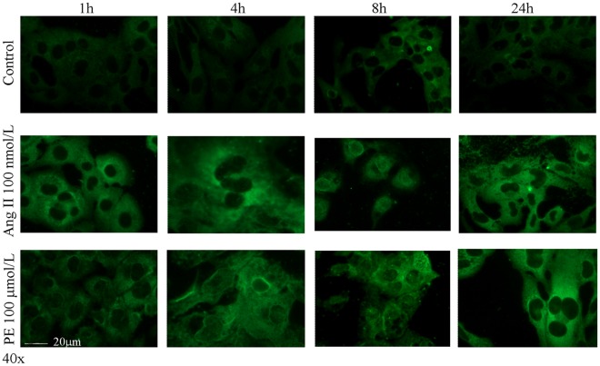
Figure 4. Immunofluorescence analysis of carbonylated proteins in cardiomyocytes after treatment with angiotensin II (AngII) or phenylephrine (PE).
The cells were stained with anti-DNP antibody and visualised by means of a secondary antibody conjugated with Alexa Fluor dye 488. Representative of three independent experiments.

