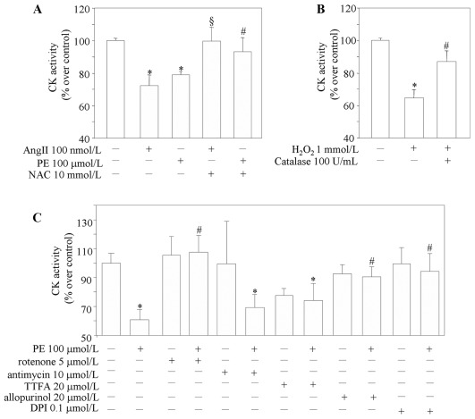
Figure 6. CK activity in cardiomyocytes.
(A) CK activity in cell lysate measured after pretreatment with NAC (N-acetyl-l-cysteine) for 1 h, and then with angiotensin II (AngII) or phenylephrine (PE) for 4 h in the absence or presence of NAC. (B) Oxidative modification of CK activity in vitro. Purified human M-CK (30 units/mL) was incubated for 1 h at 25°C with H2O2 (1 mmol/L) in 25 mmol/L Tris-HCl (pH 7.4) in the absence or presence of 100 units/mL of catalase before measurement of CK activity. *p<0.05 vs CK alone. (C) CK activity in cells lysate measured after pretreatment with different ROS inhibitors for 1 h, and then with phenylephrine (PE) for 4 h. *p<0.05 vs control; #p<0.05 vs PE-treated cells, §p<0.05 vs AngII-treated cells (n = 5).

