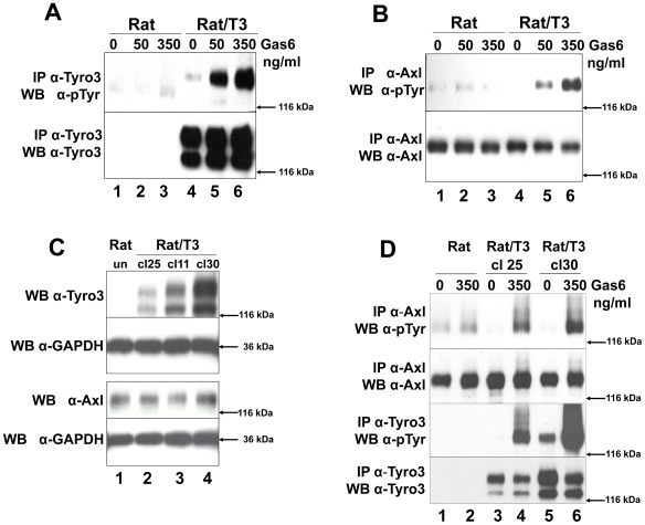
Figure 4. Tyro3 increases Gas6-induced Axl phosphorylation.
Rat2 (Rat lanes 1–3) and Rat2/T3V5 cells (Rat/T3, lanes 4–6) were treated with 0, 50, 350 ng/ml Gas6 for 20 min. Detergent cell lysates were prepared and normalized for protein concentration. The samples were divided in two for Tyro3 (panel A) and Axl (panel B) immunoprecipitations (IP). This was followed by SDS-PAGE using 8% gels followed and Western blot analysis. The membranes were probed with anti-phosphotyrosine (α-pTyr) antibodies (PY20 and P99 mixture 1∶3,500) (top, panels A and B). The membranes were stripped and reprobed with α-Tyro3 serum 5424 (1∶3,500) (A, bottom panel) or affinity purified rabbit α-Axl (1∶3,500) (B, bottom panel). These blots are representative of 4 experiments. To determine whether Tyro3 expression affects Axl levels in Rat2 cells (panel C), detergent cell extracts were prepared from Rat2 cells (lane 1), and independently derived stably transfected Rat2/T3V5 cell lines (clone (cl) 25, lane 2; clone 11, lane 3; clone 30, lane 4). SDS-PAGE using 4–20% gels followed by Western blot analysis was performed on these extracts. The membranes were cut at the level of the 66 kDa marker and the top portion was probed with rabbit α-Tyro3 (5424 serum 1∶3,500) or rabbit α-Axl (1∶3,500). The bottom portion of the membranes were blotted with α-GAPDH (1∶500). These blots are representative of 5 experiments. To determine if the levels of Axl phosphorylation depended on the levels of Tyro3 expressed (panel D) Rat2 cells (lanes 1 and 2) and Rat2/T3V5 cell lines cl25 (lanes 3 and 4) and cl30 (lanes 5 and 6) were activated with media only (0) or 350 ng/ml of Gas6 for 10 min. Detergent cell lysates were prepared and normalized for protein concentration. The samples were divided in two for Tyro3 and Axl immunoprecipitations (IP). SDS-PAGE using 8% gels followed by Western blot analysis was performed. The membranes were probed with anti-phosphotyrosine (α-pTyr) antibodies (PY20 and P99 mixture 1∶3,500) (top and third panels). An aliquot of the remaining Axl IPs were reloaded and probed with affinity purified rabbit α-Axl (1∶3,500) (second panel from the top). The membrane corresponding to Tyro3 IP’s was stripped and reprobed with α-Tyro3 serum 5424 (1∶3,500) (bottom panel). These blots are representative of 6 experiments.

