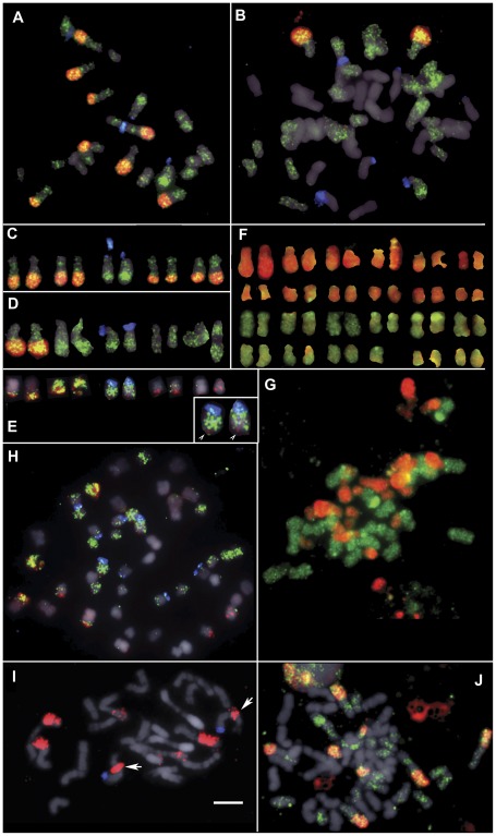
Figure 3. FISH of NicCL3 and NicCL7/30.
Fluorescence in situ hybridisation (FISH) to metaphase chromosomes of (a, c) N. tomentosiformis (ac. NIC 479/84); (b, d) Th37-3; (e, h) N. tabacum (ac. 095-55); (i) N. kawakamii and; (j) TR1-A. The probes used were 18S rDNA (blue; a-e and h-i only), NtCL7 (green) and NicCL3 probes (red) counter stained with DAPI (grey). Inset (e) shows enlarged chromosome T3 with NicCL3 signal at the distal end of the long arm (arrow heads). (f, g) Genomic in situ hybridisation (GISH) to chromosomes of Th37-3, showing the N. tomentosiformis sub-genome (red) and N. sylvestris sub-genome (green). (i) Note that chromosome 3 of N. kawakamii (18S rDNA bearing) has a large NicCL3 signal proximal to the centromere (arrows). (j) TR1-A an S0 synthetic N. tabacum with the expected number of NicCL3 (red) signals and highly localised NtCL7 (green) signals. Scale bar is 5 µm.

