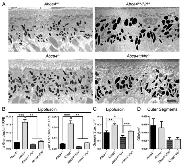
Fig. 9.
Fewer lipofuscin granules are detected in the RPE cells of ABCA4 deficient eyes in the Nrl−/− background when compared to the WT background. (A) Representative electron micrographs of the RPE cell layer at P120 showing lipofuscin granules (black arrows) and phagocytosed outer segments (white arrows). Scale bar 2 μm. For quantification, 3–5 animals per group were analyzed and at least 5 images per eye were counted, totals were summed for each eye and values shown are means±SEM. (B) Both the number of lipofuscin granules per μm of RPE area (left) and the fraction of RPE occupied by lipofuscin granules (right) are increased with significance in the Abca4−/− compared to all other genotypes, and these parameters are also non-significantly increased in Abca4−/−/Nrl−/− compared to Abca4+/+/Nrl−/−. (C) In both the WT and Nrl−/− backgrounds, the average size of each lipofuscin granule is larger in the absence of ABCA4. (D) In the Nrl−/− background the number of OS pieces detected inside the RPE appeared to be less than in the WT background, but the difference was not statistically significant. *p<0.05, **p<0.01, ***p<0.001.

