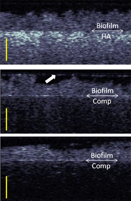
Figure 3.
Cross-Polarization OCT images of the microcosms after 48 hours of growth on (top) hydroxyapatite, (middle) silorane based composite, (bottom) methacrylate based composite. The double arrow demarcation shows the boundary between the biofilm and the material. The scale bars are 500 μm in optical depth or roughly ~385 μm in biofilm depth. The arrow (middle image) shows a highly reflecting water layer that causes some crosstalk into the cross polarization channel.

