
Figure 11.
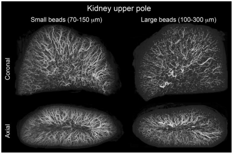
microCT of swine kidney tissue embolized with radiopaque DEBs. Small size range (70–150 μm) and large size range (100–300 μm) DEBs are displayed with consistent size scaling. Small beads penetrate to more distal regions and yield a greater spatial density. These DEBs were made radiopaque by the inclusion of Lipiodol inside the DEBs. From [77] with permission.