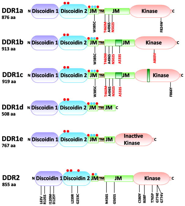
Fig. 1.
Domain structure of DDRs. Residues that are added as a result of alternative splicing are indicated by dark green boxes within the corresponding domain. Red and blue circles indicate putative N-glycosylation and O-glycosylation sites, respectively. Mutated DDR residues, which were identified in samples of Non-Small Cell Lung Carcinomas (black) and Acute Myeloid Leukemia (red), are indicated. aa, amino acids; JM, juxtamembrane region, and TM, transmembrane region.

