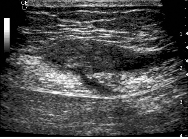
Figure 3.

A 30-year-old woman with a long history (84 mo) of continuous pain in the lower abdomen and two previous non-diagnostic laparoscopic examinations. In the abdominal wall (between the subcutaneous fat and the muscle), US exam discloses a 4-cm, ovoid hypoechoic endometrioma with a linear fistulous tract (arrow) emerging from the posterior aspect of the lesion and transgressing the muscular plane.
