
Figure 3.
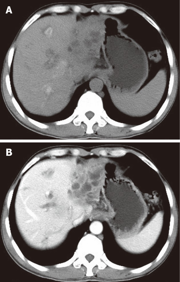
A 54-year-old male with liver malignant fibrous histiocytoma. A: Non-contrast computed tomography shows ill defined lesion of density 30 HU in the left lobe with dilated intrahepatic biliary duct; B: Contrast scan portal vein phase shows low enhancement compared to normal liver parenchyma and multiple areas of low attenuation.