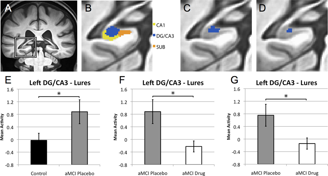
Figure 2. Levetiracetam treatment normalizes increased dentate gyrus/CA3 activity in patients with aMCI.
(A) High-resolution anatomical scan, with the left medial temporal lobe demarcated. (B) Segmentation of medial temporal lobe structures, including the CA1, dentate gyrus/CA3 (DG/CA3) and subiculum (SUB) subregions of the hippocampal formation. The boundaries between the CA3 and dentate gyrus (DG) cannot be clearly defined in structural scans and, therefore, are combined as DG/CA3 for analysis. (C) Area of task related activity based on an ANOVA of task condition in the left DG/CA3. (D) Area of task related activity in the left DG/CA3 resulting from the confirmatory analysis in which voxel selection is based on ANOVA of task condition that includes only age-matched healthy control subjects. (E) Graphs show mean activity ± SEM on lure trials correctly called similar. Patients with aMCI on placebo show increased activity in the left DG/CA3 compared to healthy control subjects during the lure trials. (F) Levetiracetam treatment significantly reduced activity in the DG/CA3 in patients with aMCI compared to the placebo condition. (G) In the confirmatory analysis for drug treatment, patients with aMCI taking low dose levetiracetam again showed significantly reduced BOLD activation during lure trials correctly identified as similar relative to the activity during those trials under placebo treatment. * p < 0.05 group comparisons using independent and paired samples t-tests respectively. See also Figure S1.

