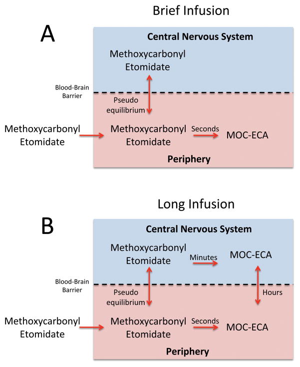
Figure 7.
Schematic illustration of our current theory regarding the relationships between methoxycarbonyl etomidate infusion time, post-infusion recovery time, methoxycarbonyl etomidate carboxylic acid (MOC-ECA) concentrations in the blood and cerebrospinal fluid, and the rate of methoxycarbonyl etomidate metabolism in brain tissue and cerebrospinal fluid in the rat. The approximate time scale for each process is given. (A) With brief methoxycarbonyl etomidate infusion (or single bolus), there is insufficient time for methoxycarbonyl etomidate to be metabolized to MOC-ECA within the central nervous system or for metabolite formed in the blood to diffuse into the central nervous system. Consequently, there is little MOC-ECA in the brain and essentially all of the burst suppression and hypnosis is produced by methoxycarbonyl etomidate alone. Upon terminating the infusion, the methoxycarbonyl etomidate concentration in the brain falls rapidly in parallel with that in the blood because methoxycarbonyl etomidate diffusion across the blood-brain barrier is very fast. Recovery is rate-limited by peripheral (e.g. blood) metabolism of methoxycarbonyl etomidate, which occurs on the time scale of seconds. (B) With longer infusions, significant quantities of MOC-ECA are formed from methoxycarbonyl etomidate within the central nervous system. This metabolic process occurs on the time scale of minutes. With sufficiently long infusions, essentially all of the burst suppression and hypnosis is produced by MOC-ECA. Upon terminating the infusion, the MOC-ECA concentration in the brain falls slowly because it crosses the blood-brain barrier and ultimately excreted from the body on the time scale of hours.

