Figure 1.
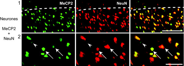
All neurones express MeCP2 in the rat superficial dorsal horn. Confocal images of rat superficial dorsal horn sections. Colocalization of MeCP2 (green; Millipore antibody) and NeuN (red). MeCP2 can be seen within the nucleus of all neurones (arrows). However, some MeCP2 staining is clearly non-neuronal (arrow heads). Pictures show single focal plane. Scale bars, 1) 50 μm and 2) 20 μm.

