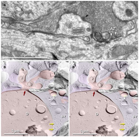
Figure 9.
Thin-section (A) and freeze-fracture images (B) of thorny excrescences, with lateral extension of MF terminal forming glutamatergic synapses onto dendritic shafts of CA3pyr. Tightly clustered synaptic vesicles are at asymmetric synapses, occasionally with electron-dense presynaptic active zones [(A); white arrowheads opposite dendritic shaft; black arrowhead opposite spine]. (B) FRIL image of cross-fractured dendrite shaft, with thorny excrescence extending into the complex spine apparatus. (Base of spine indicated by red arrow.) One PSD (yellow overlay) with a single gold bead (white arrowhead) is labeled for NMDA-R1. A portion of a complex MF terminal (purple overlay) is traceable from an included spine to its contact with the cross-fractured CA3pyr dendrite (D; red overlay). In the apposed, MF terminal, synaptic vesicles (sv) are densely clustered at probable active zones (yellow arrows). Scale bars are 1 μm.

