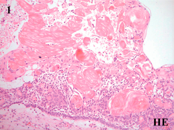
Figure 1.

Microscopic features of the calcifying cystic odontogenic tumor showing fibrous connective tissue wall lined by ameloblastomatous epithelium characterized by basal low columnar/cuboidal cells and suprabasal stellate reticulum-like cells with interspersed clusters of ghost cells (Hematoxylin & eosin, ×100).
