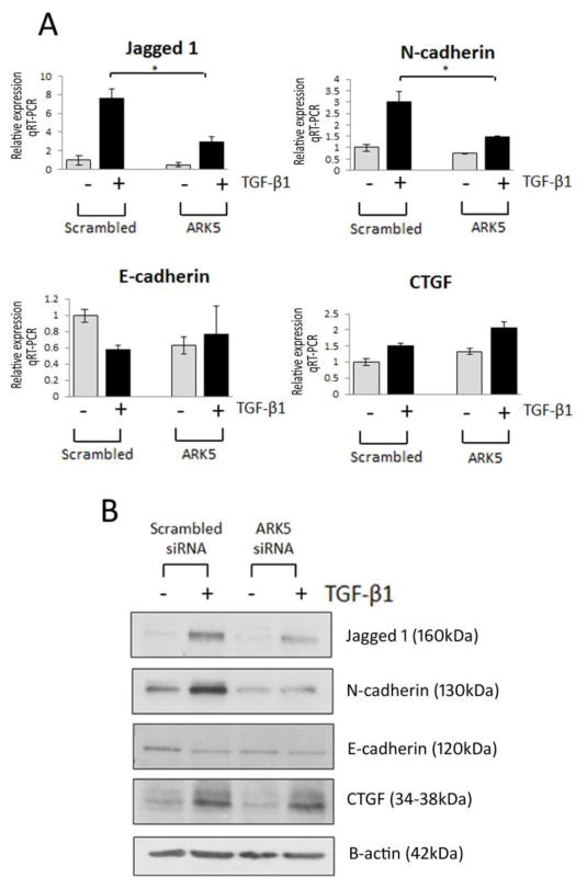
Fig. 6.
Role for ARK5 in TGF-β1-induced fibrosis in renal epithelial cells. (A) TaqMan quantitative PCR analysis of Jagged1, N-cadherin (CDH2), E-cadherin (CDH1) and CTGF in HK-2 cells transfected with ARK5 siRNA in the absence (grey bar)/presence (black bar) of TGF-β1 (5ng/ml; 48h). (B) Representative Western blot of Jagged1, N-cadherin (CDH2), E-cadherin (CDH1) and CTGF HK-2 cells transfected with ARK5 siRNA, respectively. HK-2 cells transfected with scrambled siRNA were selected as a control. For TaqMan PCR, expression was normalized to GAPDH. Data are plotted as mean ± SE. *P < 0.05, Student’s unpaired t test, n = 3 per group.

