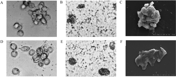
Figure 1.

Morphology of rhesus macaque monocyte-derived DC. (A) Cell colonies of dendritic cell precursors of antigen-untreated group on day 3 of culture (magnification, × 400). (B) Morphology of DC of inactivated SV40-treated group on day 9 of culture (magnification, × 400). (C) Morphology of DC of inactivated SV40-treated group on day 12 of culture (magnification, × 2500). (D) Cell colonies of dendritic cell precursors of antigen-untreated group on day 9 of culture (magnification, × 400). (E) Morphology of DC of infective SV40-treated group on day 9 of culture (magnification, × 400). (F) Morphology of DC of infective SV40-treated group on day 12 of culture (magnification, × 1200).
