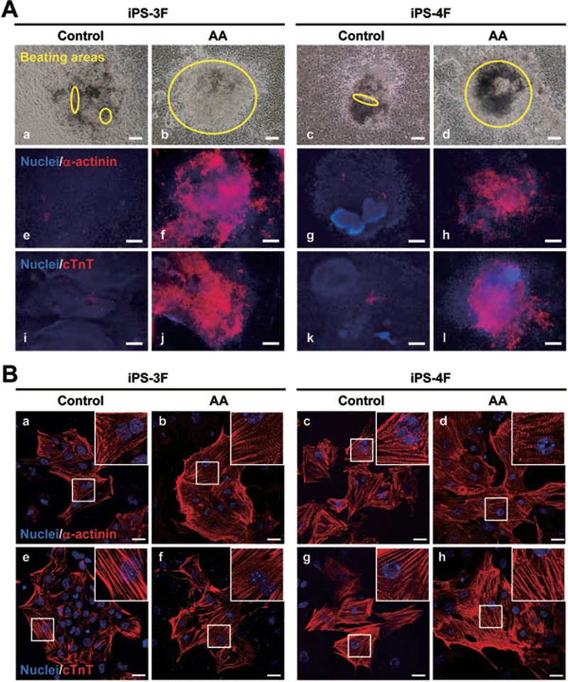
Figure 2.
AA increases the content and improves the sarcomeric organization of iPS-CMs. (A) Representative images showing increased beating areas (a-d) and the content of α-actinin+ (e-h) or cTnT+ (i-l) cardiomyocytes in day-10 EBs treated with AA. Scale bars = 100 μm. (B) Sarcomeric structure analysis of the day-18 iPS-CMs by α-actinin and cTnT staining. The insets were magnifications of the framed areas showing more organized cross-striation alignment of sarcomeres in AA-induced iPS-CMs. Scale bars = 25 μm. Nuclei were counterstained with Hoechst33258 (blue).

