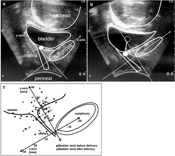
Figure 1a+b.

Perineal ultrasound for evaluating the bladder neck mobility. Bladder neck at rest (Figure 1a) and with valsalva (Figure 1b), point indicates the bladder neck, arrow as vector for the bladder neck mobility. Figure 1c: Position of the bladder neck before and after delivery at rest.
