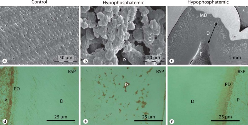
Fig. 1.
Dentin examination by SEM and IHC (anti-BSP) of a control molar (a, d), a hypophosphatemic primary molar (b, e) and a hypophosphatemic permanent molar (c, f). The inner part of dentin appears normal in the permanent hypophosphatemic molar, whereas discrete calcospherites are observed close to the mantle dentin. P = Pulp; PD = predentin; D = dentin; MD = mantle dentin; C = calcospherite; IS = interglobular space.

