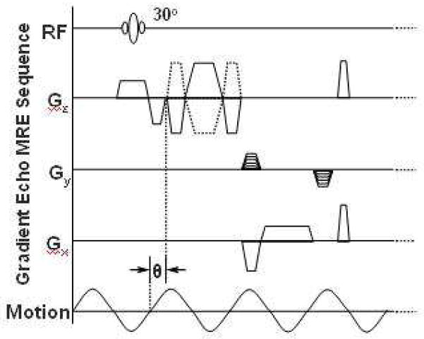
Figure 2.
MRE pulse sequence diagram illustrating the timing of the imaging and motion-encoding gradients in relation to the applied vibration. Theta indicates an adjustable phase delay between the motion and the motion-encoding gradients to capture the tissue motion at different time points during the wave propagation.

