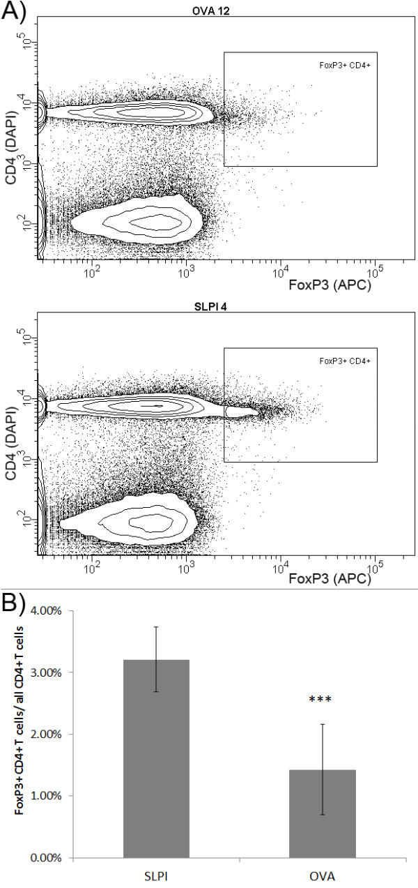
Figure 5.

SLPI protein immunization reduces number of FoxP3+ CD4+ T cells in lymph nodes of in vivo. A: 10 OVA- and 9 SLPI-immunized SJL/J were sacrificed 12 days after passive EAE induction. LNC were extracted and analyzed by flow cytometry for CD4 (DAPI channel) and FoxP3 (APC channel). Result from a representative OVA-immunized mouse is shown in the upper diagram and from a SLPI-immunized mouse in the lower diagram. B: Column chart represents the average ratio of FoxP3+ CD4+ and total number of CD4+ T cells in inguinal lymph nodes of SLPI- and OVA-immunized mice on day 12 after passive EAE induction (***, p < 0.001, Student's t-test)
