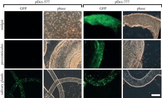
Figure 5.
Inducible transgene expression in SmOxP927 cells in different compartments of the tsetse fly. Forty-eight hours-induced cells are shown for both the pDex-577 and pDex-777 plasmid. All images taken at the same magnification; scale bar indicates 50 μm. Representative GFP fluorescence (GFP) and phase contrast (Phase) images are shown for each infected compartment of the fly. The midgut image for pDex-577 shows trypanosome cells that have spilled out of the dissected midgut. The midgut image for pDex-777 shows a midgut full of fluorescent trypanosome cells, in which it is difficult to resolve individual cells. The proventriculus images for both pDex-577 and pDex-777 show an infected proventriculus with trypanosome cells. The salivary gland images for pDex-577 and pDex-777 show an infected salivary gland.

