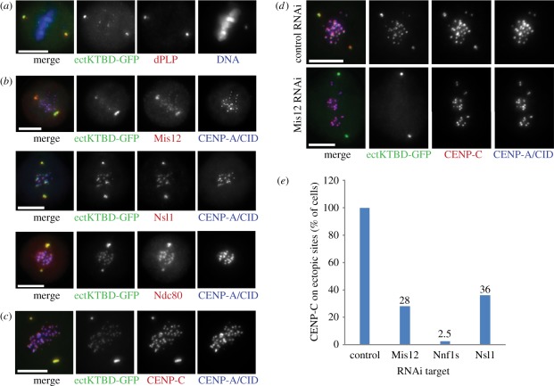
Figure 5.
Ectopic expression of the kinetochore-binding domain of Spc105 fused with EGFP (ectKTBD-GFP). (a) ectKTBD-GFP (green) colocalizes with dPLP (red) in Dmel-2 cells. DNA counterstained with DAPI (blue). (b) KMN network components (red) Mis12, Nsl1 and Ndc80 are recruited to centrosomes in cells expressing ectKTBD-GFP (green). CENP-A/CID (blue) signals mark the endogenous centromeres; CENP-A/CID is not recruited to ectopic sites. (c) CENP-C (red) localizes to endogenous centromeres and to ectopic sites, just like ectKTBD-GFP (green). (d) Negative control after RNAi shows the pattern of staining identical to the one shown in (c). However, the depletion of Mis12 complex components (here Mis12 protein is given as an example) displaces ectKTBD-GFP from endogenous centromeres and CENP-C from ectopic sites. (e) After RNAi, described in (d), cells showing the ectopic CENP-C localization were counted (n = 40 in each case). All ectKTBD-GFP expressing cells show ectopic CENP-C in a control knock down, but CENP-C is retained on centrosomes only in a fraction of cells after depletion of the individual Mis12 complex subunits. Scale bars, 10 µm.

