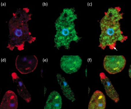
Fig. 5.
Confocal micrographs of N. fowleri and N. lovaniensis placed on collagen I. N. fowleri was probed with Alexafluor 594 phalloidin (a) or FITC-conjugated monoclonal anti-β1 integrin antibody (b). The merged fluorescent image is shown in (c). N. lovaniensis was probed with Alexafluor 594 phalloidin (d) or FITC-conjugated monoclonal anti-β1 integrin antibody (e). The merged fluorescent image is shown in (f). Nuclear localization is depicted in all panels by DAPI staining (a–f). All images are magnified ×100.

