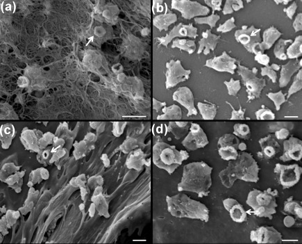
Fig. 8.
Interaction (2 h) of N. fowleri and N. lovaniensis with collagen I and Matrigel matrices. (a) N. fowleri emerging through the bottom side of the collagen I scaffold and (b) N. lovaniensis interacting with collagen I on the surface of the scaffold. (c) N. fowleri interacting with the Matrigel surface. (d) N. lovaniensis interacting with the Matrigel surface. Note the presence of ‘food-cups’ (arrows) on both N. fowleri and N. lovaniensis. Bars, 10 µm.

