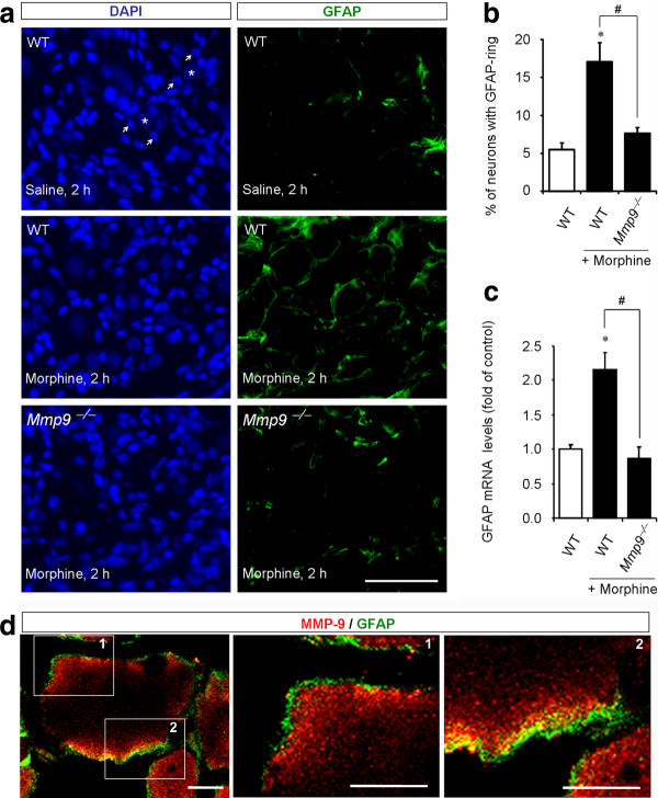
Figure 3.
MMP-9 is required for subcutaneous morphine-induced GFAP expression in SGCs of DRGs. (A) Left panels: DAPI nuclei staining of all cells in DRG sections of saline (up panel) and morphine (2 h, middle and low panels) treated wild-type (WT) mice and morphine-treated Mmp9 knockout mice (Mmp9-/-, low panel). Stars and arrows indicate neurons (no staining) and SGCs, respectively. Note that Mmp9 knockout mice show no changes in DRG cell numbers. Right panels: Immunohistochemistry showing GFAP-labeled SGCs in DAPI-stained DRG sections. Scale, 50 μm. (B) Percentage of DRG neurons with GFAP-labeled ring. Note that morphine-induced GFAP increase at 2 h is abrogated in Mmp9 knockout mice. *P < 0.05, compared to WT control; #P < 0.05 compared to morphine/WT, ANOVA followed by Bonferroni post hoc test, n = 6 mice. (C) GFAP mRNA expression in DRGs revealed by real time RT-PCR analysis. Note that morphine-induced GFAP mRNA expression (2 h) in WT mice is abolished in Mmp9 knockout mice. *P < 0.05, compared to WT control; #P < 0.05 compared to morphine/WT; ANOVA followed by Bonferroni post hoc test, n = 4. (D) Confocal images showing doubles staining of MMP-9 and GFAP in DRG sections of WT mice 2 h after morphine treatment. Square 1 and 2 are enlarged in middle and right panels. Note there is close proximity but not overlap of MMP-9 and GFAP staining. Scales, 10 μm.

