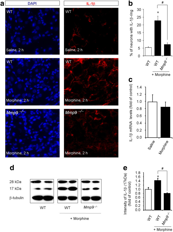
Figure 4.
MMP-9 is required for subcutaneous morphine-induced IL-1β activation in DRGs. (A) DAPI nuclei staining (left panels) and IL-1β immunostaining (right panels) in DRG sections of saline (upper panels) and morphine (middle panels) treated wild-type (WT) mice and morphine-treated Mmp9 knockout mice (Mmp9-/-, lower panels). Scale, 50 μm. (B) Percentage of DRG neurons with IL-1β-labeled ring. Note that morphine-induced IL-1β increase is abrogated in the Mmp9 knockout mice. *P < 0.05, compared to WT control; # P < 0.05 compared to morphine/WT, ANOVA followed by Bonferroni post hoc test, n = 6 mice. (C) Real-time RT-PCR analysis showing IL-1β mRNA expression in DRGs of saline and morphine treated mice (10 mg/kg, s.c., 2 h). P > 0.05, Mann-Whitney test, n = 4. (D) Western blotting showing non-mature (28 kDa) and mature/active (17 kDa) bands of IL-1β in DRGs of WT and Mmp9-/- mice with or without morphine treatment (10 mg/kg, s.c., 2 h). Note that the 28 kDa bands were unaltered after morphine treatment. (E) Intensity of the active IL-1β (17 kDa) bands. Note that morphine-induced activation of IL-1β is abrogated in Mmp9-/-mice. *P <0.05, compared to WT control; #P <0.05 compared to morphine/WT, ANOVA followed by Bonferroni post hoc test, n = 4 mice.

