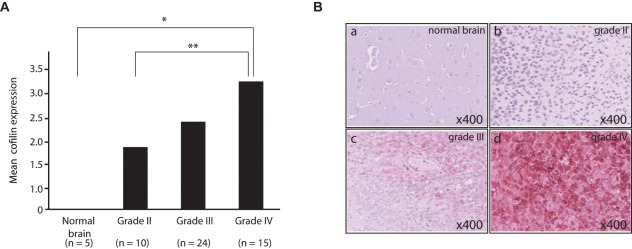
Figure 2.
Increased cofilin expression correlates with increasing grade malignant astrocytoma. (A) Results of cofilin immunohistochemistry on brain tumor tissue microarray represented as a bar graph showing distributions of total score of immunostaining for cofilin in each group according to histopathological grade of astrocytoma. Total scores were determined by both distribution within a section and staining intensity. Asterisks indicate statistically significant differences (*P = 0.004, **P = 0.034). (B) Images of representative specimens immunostained for cofilin from each group. The original histopathological diagnoses are normal brain (a), diffuse astrocytoma (grade II; b), anaplastic astrocytoma (grade III; c), and glioblastoma (grade IV; d) Original magnification 200×.

