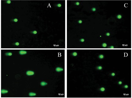
Figure 5.
DNA damage assessed by comet assay (representative micrographs of fluorescent DNA stain of A549 cells are shown). (A) Untreated cells with undamaged DNA (negative control). (B) Cells exposed to 100 mM of H2O2 for 20 minutes at 4°C (positive control; denatured DNA fragments migrate out from the nucleoid in a different length of comet tail). (C, D) FDH-expressed cells have undamaged supercoiled DNA (it remains inside of the nucleoid). Cells were examined at different time points (C: 24 hours; D: 48 hours) of FDH induction.

