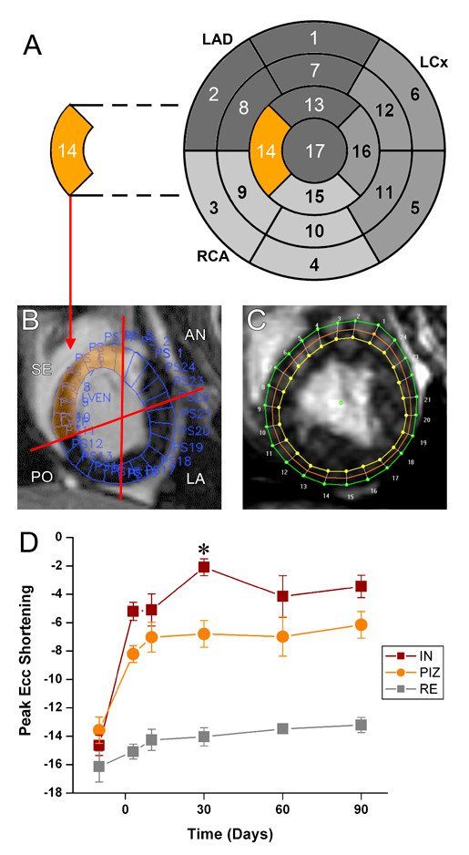
Figure 5.

Strain analysis in the peri-infarct zone in short axis slices. (A) The 17-segment analysis provides an overview regional function in the whole left ventricle. (B) However, data from the infarct, the PIZ and non-infarcted areas have to be combined, and are therefore averaged. The septal (SE) area highlighted in orange represents segment 14. (SE = septal; AN = anterior; LA = lateral; PO = posterior). (C,D) To assess regional strain in the PIZ we sub-divided the short axis slices in 24 segments and matched each segment with the corresponding segment from the tagging analysis for Ecc and Err analysis, respectively.(D) At 30 days post-MI peak Ecc is different between the infarct scar tissue (IN) and the PIZ (* p < 0.05 versus IN). (F) At 3 days and 30 days post-MI the Err shows different strain values in the IN and PIZ(* p < 0.05 versus IN). (RE = remote moyocardium, PIZ = peri-infarct zone, IN = myocardial infarct scar).
