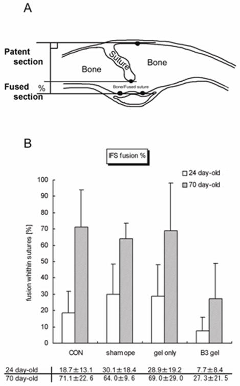
FIG. 1.
A, Diagram showing a coronal section through the calvaria with a partially endocranially fused interfrontal suture (IFS) between left and right frontal bones. The thickness of the bones was measured, and the distance between top and bottom horizontal line gives the total width of the suture. Sutures begin fusing from the endocranial surface, so the distance between the middle and bottom horizontal lines gives the width of the fused region of the suture. This scoring method assessed the degree of sutural fusion across the thickness of the bones to test the effect of Tgf-β3 or lack of it. This measurement was not meant to correlate suture morphology (such as interdigitation, beveling, and fusion) and biomechanics, which requires a three dimensional image database. B, Graph showing the percent fusion of sutures in each group.

