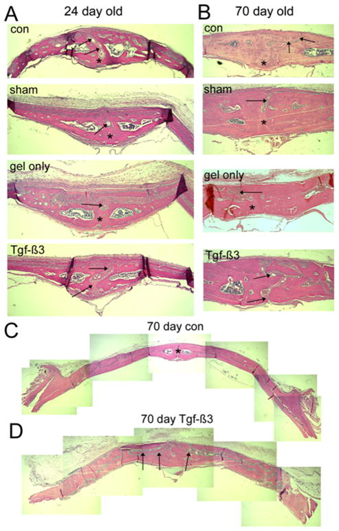
FIG. 3.
Photomicrographs of histological sections through the posterior IFS of 24 and 70 day old animals. The ectocranial (periosteal) surface is superior and endocranial (dural) surface is inferior in all micrographs. Frontal bones are to the left and right of the suture (arrows). A, IFS of 24-day animals, showing sutures (arrows) in the control (con), sham operated (sham), and Gel only groups with bony bridging on their dural aspect (asterisks) by 24 days. In all cases the sutures fused from the dura side. Sutures in the Tgf-β3 treated group remained open (arrows). B, IFS of 70-day animals, showing gradual thickening of the bones compared to the 24-day animals. Note the progressive bony bridging (asterisks) across the 70-day control, sham and gel only sutures (arrows), with the persistent patency of the Tgf-β3 treated IFS (arrows). C, Low power composite images of sections through calvariae of control and Tgf-β3 treated animals. Note the thicker bones in the Tgf-β3 treated calvaria, with the highly complex IFS (arrows) winding through the bone.

