
Fig. 3.
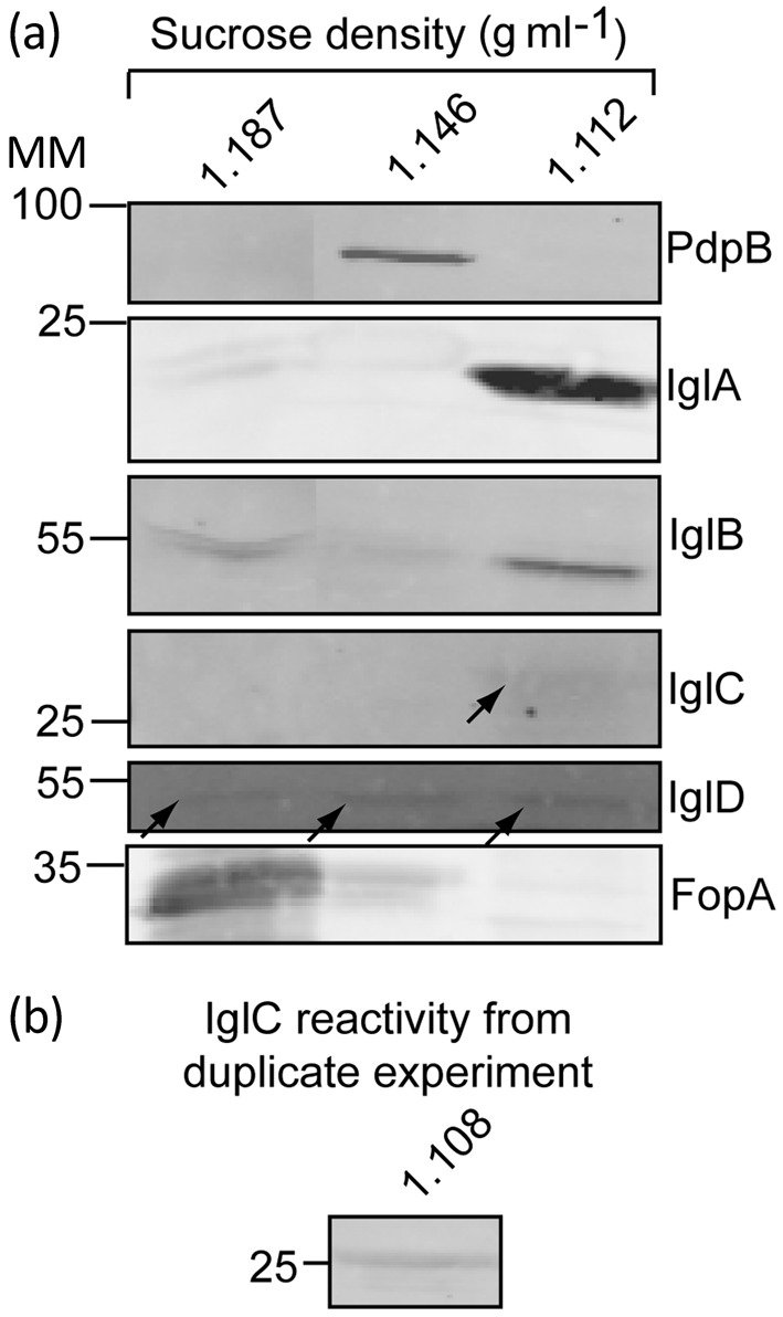
Localization of IglA, IglB, IglC and IglD following osmotic lysis of F. tularensis LVS cells. (a) Immunoblot analysis of fractions from sucrose gradient separation of insoluble material from a bacterial extract generated by osmotic lysis of cells. (b) Immunoblot of IglC from a similar sucrose fraction from a duplicate experiment of that shown in (a). MM, molecular mass (kDa).