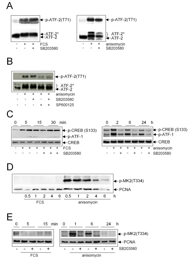
Figure 4.
Analysis of p38-mediated substrate phosphorylation. (A) Serum-starved NIH3T3 cells were either not pretreated or pretreated with SB203580. Cells were not stimulated, stimulated with FCS (left) or with anisomycin (right) for 15 min. Total cell extracts were performed and subjected to Western blot analysis. Phosphorylation of ATF-2 was detected using a phospho-specific antibody, equal loading was controlled by stripping and reprobing the blot with anti-ATF-2-antibody. (B) NIH3T3 cells were either not treated or treated with anisomycin in the absence or presence of SB203580 (10 μM), SP600125 (25 μM) or both inhibitors for 15 min. Western blot analysis was performed as described above. (C) Serum-starved NIH3T3 cells were either not pretreated or pretreated with SB203580 and then stimulated with FCS (left) or anisomycin (right) for the indicated time points. Total cell extracts were performed and subjected to Western blot analysis. Phosphorylation of CREB was detected using a phospho-specific antibody, equal loading was controlled by stripping and reprobing the blot with anti-CREB-antibody. The anti-phospho-CREB-antibody also recognizes phosphorylated ATF-1. ATF-2* = phosphorylated ATF-2. (D) Serum-starved NIH3T3 cells were either not treated or treated with FCS or anisomycin for the indicated time points. Western blot analysis was performed using a phospho-specific anti-MK2-antibody. The blot was stripped and reprobed with anti-PCNA-antibody to control equal loading. (E) Serum-starved NIH3T3 cells were either not treated or treated with FCS or anisomycin for the indicated time points in the absence or presence of SB203580 (10 μM). Western blot analysis was performed using a phospho-specific anti-MK2-antibody. The blot was stripped and reprobed with anti-PCNA-antibody to control equal loading.

