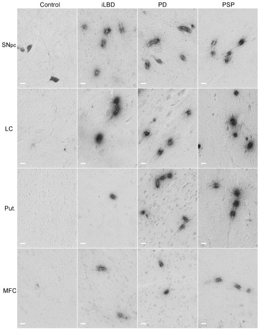
Fig 1. Integrin αvβ3 staining in post-mortem human brain tissue.
Endothelial cells of human post-mortem brain tissue were labeled with mouse anti-human integrin αvβ3 antibody and visualized with the chromagen DAB. Integrin αvβ3 reactive vessels are shown in post-mortem tissues from non-pathological controls, incidental Lewy Body Disease (iLBD), Parkinson’s disease (PD) and progressive supranuclear palsy (PSP) subjects. Note the distinct pattern of staining along vessels. In most cases only a small portion of the longitudinal vessel is stained. In other cases the vessels are perpendicular resulting in a ring of staining. The grey cells seen in the SNpc are neuromelanin-containing cells that are evident in unstained tissues and could be distinguished from the αvβ3 staining by stain color (in the original color images), and by morphology. Black scale bars = 100 μm.

