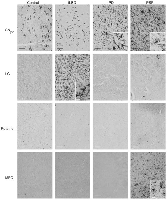
Fig 4. Activated microglia in post-mortem human tissue samples.
Activated microglia were labeled using an antibody to MHC class II antigen and visualized with the chromagen DAB. In the SNpc, there were neuromelanin-containing cells, as evident in the control condition. Microglia could be distinguished form neuromelanin containing cells by size and morphology. The black scale bar = 100 μm. Insets in the panels were taken at higher magnification and show microglia morphology typical of the microglia in that condition. White scale bars in the insets are 20 μm.

